Applications by Specialty
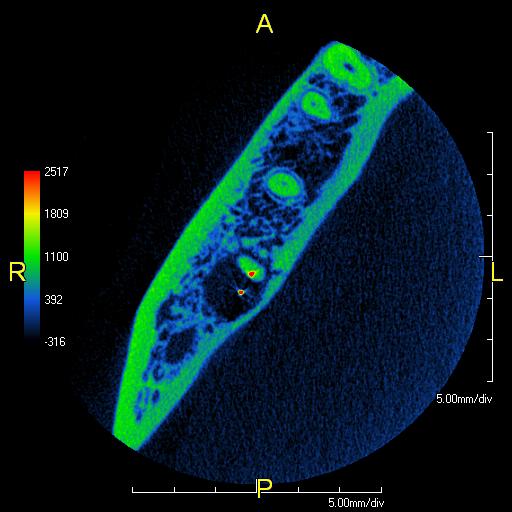
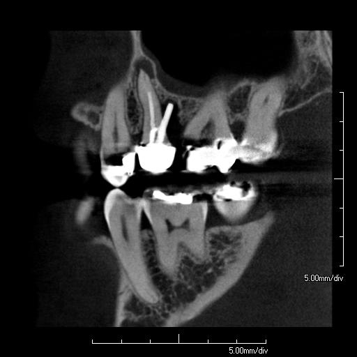
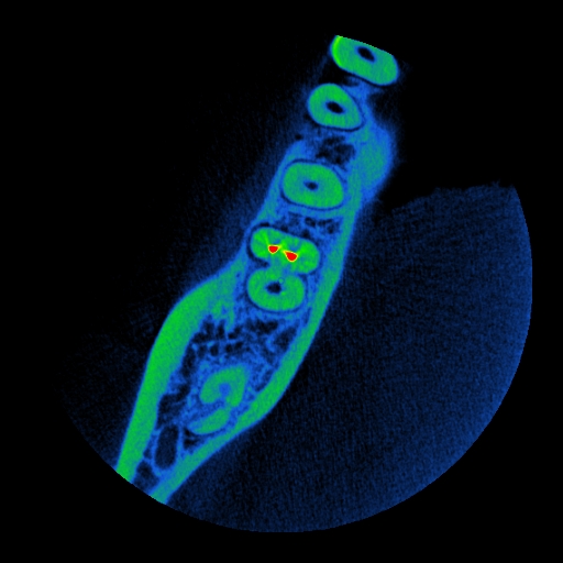
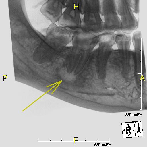
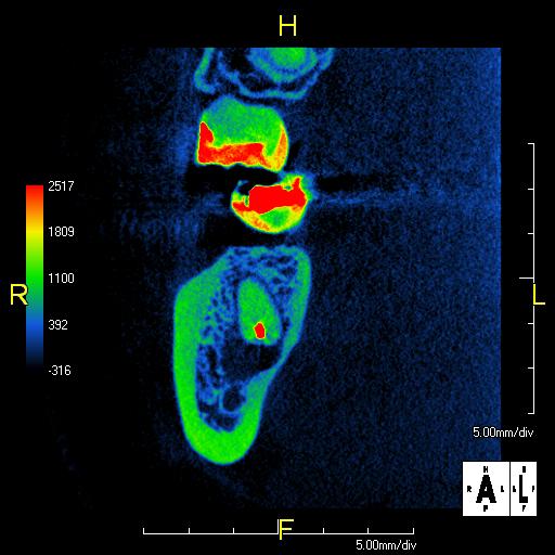
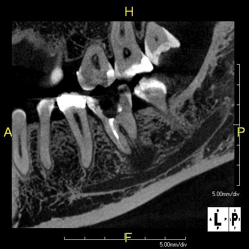
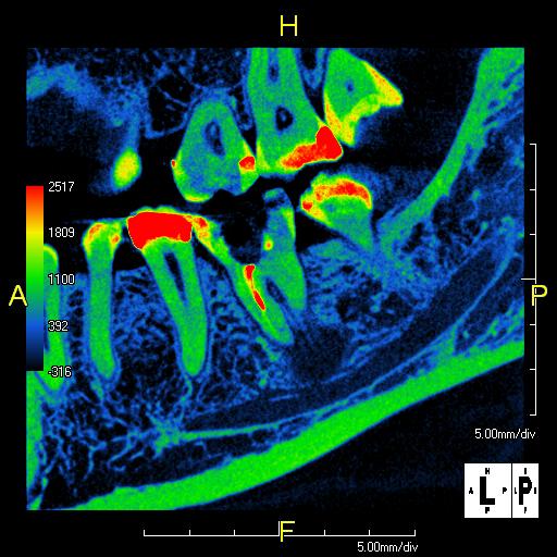
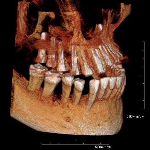
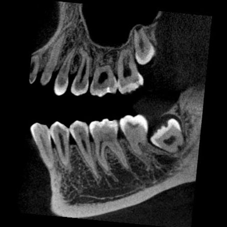
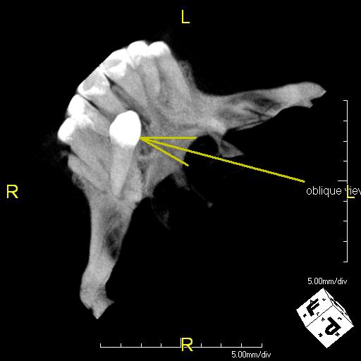
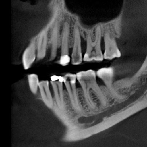
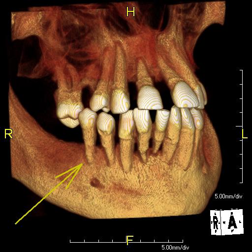
Utilizing PreXion3D CBCT will change the way you view endodontics from the initial diagnosis to retreatments. The ability to look at a tooth from virtually any angle eliminates surprises. The high-quality images show MB2 canal, split root systems, multiple root exits, and make treatment success more certain.
PreXion helps to:
- Reduce or eliminate endodontic retreatment rates by having more complete information
- Determine exact location of potential second Mesial-Buccal canals prior to surgery
- Accurately determine risk/benefit of retreatment or tooth extraction
- Clearly diagnose vertical root fractures, retained root tips, apical radiolucency
- See the potential impact of an abscess on surrounding structures
- Detect internal or external resorption
What Endodontists Are Saying…
“PreXion 3D’s clarity, resolution and unique 3D templates blow me away! For diagnostics, endo, perio, restorative and implants I don’t know how I was able to practice great dentistry without it. The customer service is truly superb and knowledgeable. Don’t walk, run and get this machine. It will make you money and give your patients the best dentistry has to offer.”
“This machine is great. Images are clear. I love it.”
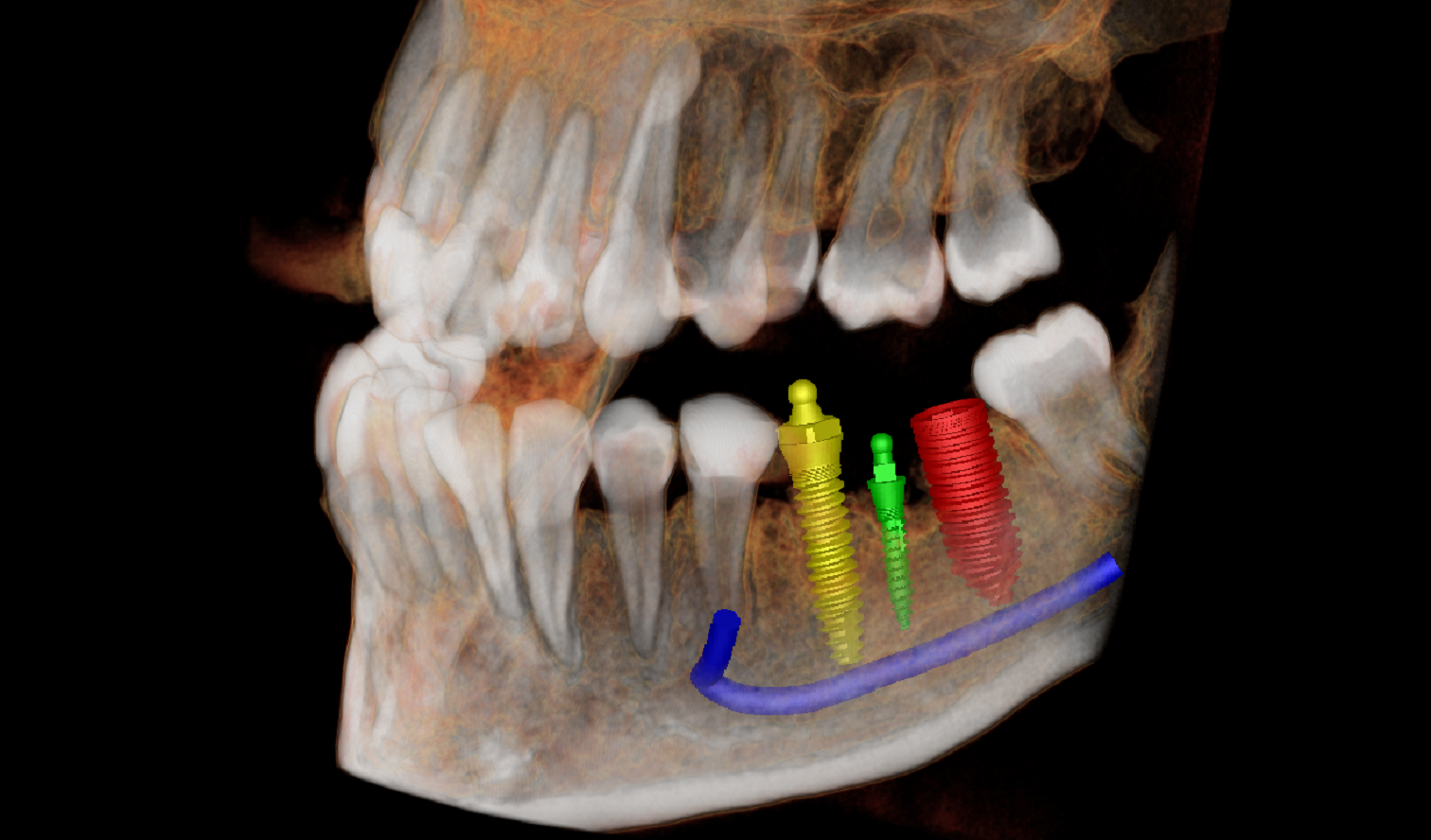
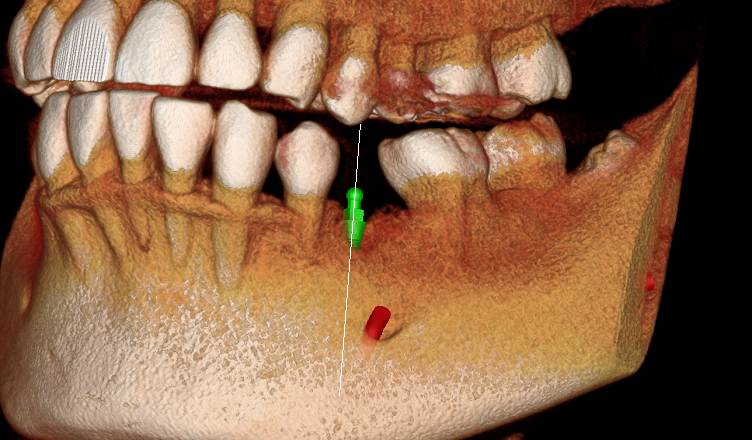
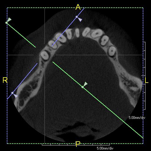
PreXion provides:
- Measurements that are exact 1:1 ratio with no superimposed structures or magnification
- The ability to assess detailed bone quality and quantity and quickly determine if grafting is required
- The opportunity to eliminate contingency treatment plans and surprises during implant surgery
- Less surgical trauma or exploratory surgery for patients
- An increase in patient education and case acceptance
- Quality 3D scan data in order to create optimal surgical guide fit
- Better implant site selection and more predictable prosthetic outcomes
What Implant Dentists Are Saying…
“I spent a lot of time looking into the purchase of my first 3D imaging system for my periodontal and dental implant practice. I chose PreXion for the image quality and outstanding software to view the data. It is amazing how quality 3D imaging will change your ability to offer the patient comprehensive periodontal and implant treatment plans with complete confidence. If you have the PreXion 3D images available at the treatment planning appointment your patient is better informed and case acceptance increases.”
“We looked at many options for our CT scanner and found PreXion to be our best choice. I cannot imagine not having the capability it gives us for specific planning for block grafts, sinus grafts and implant placement! In addition, when we show the images and our planning capability to patients, they are immediately impressed with our advanced technology and case acceptance is automatic.”
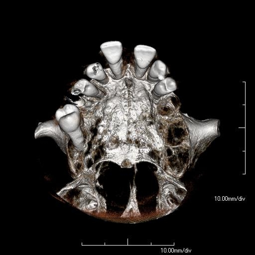
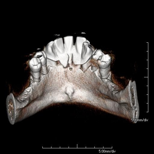
PreXion allows for:
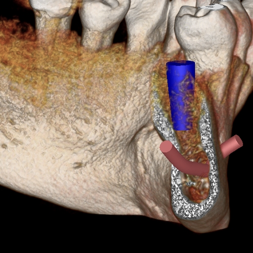
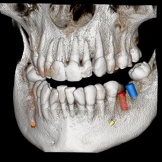
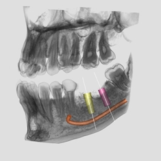
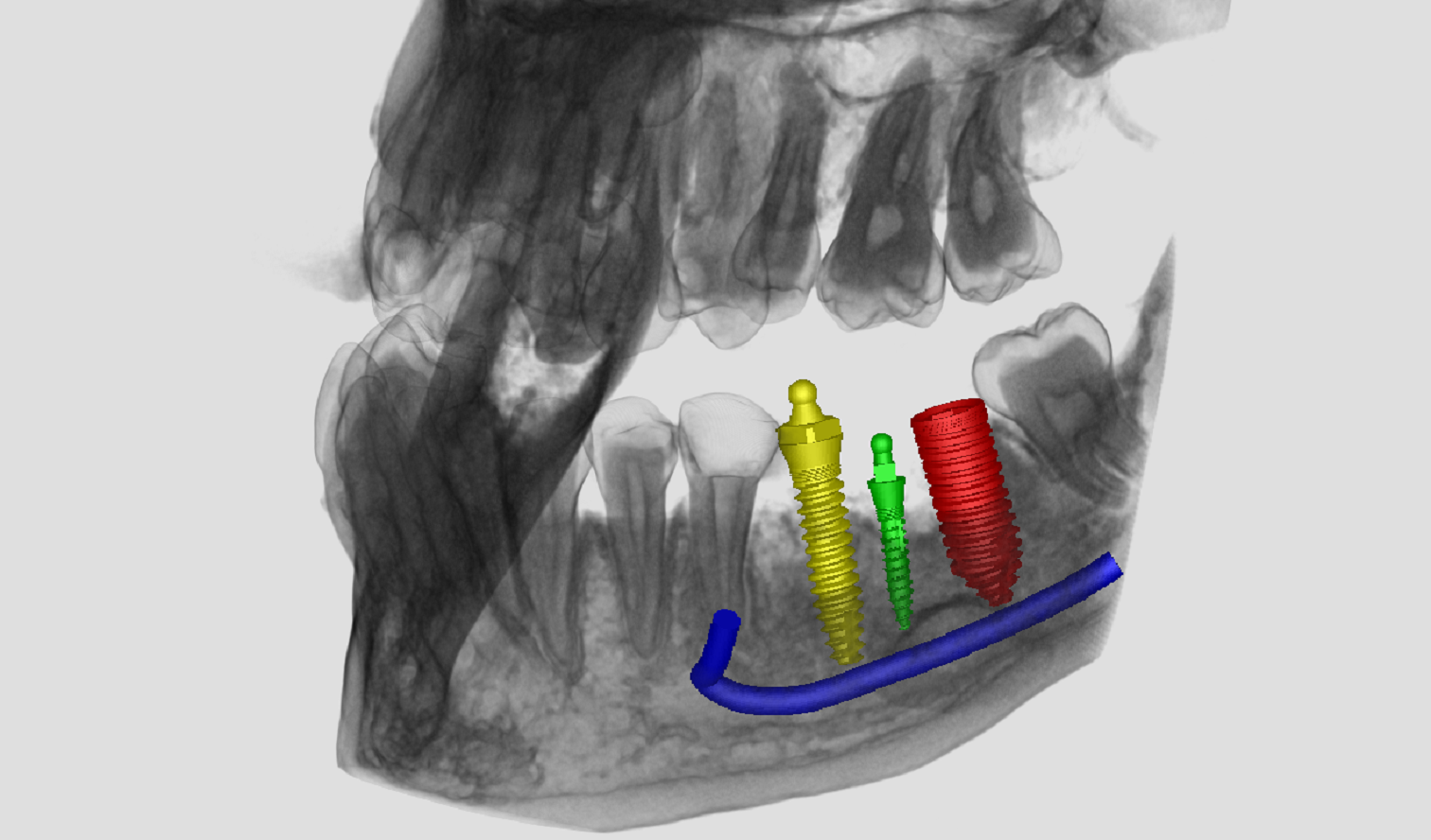
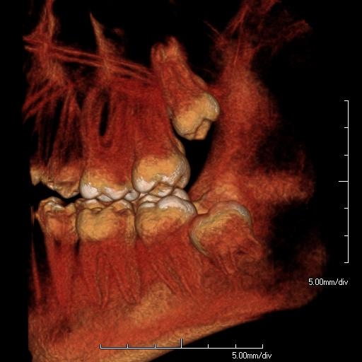
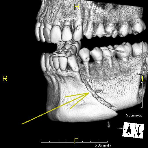
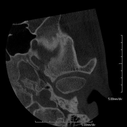
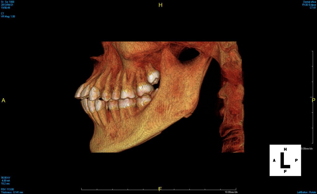
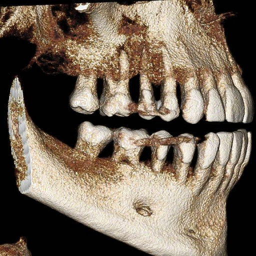
Impacted Teeth and Third Molar Studies
- Accurate evaluation of impacted third molars for proximity, angle and tooth roots
- Elimination of guesswork of third molar roots relative to Inferior Alveolar Nerve canal
- Rotational and multi-slice features that show true position of deep or curved roots
- The ability to better manage impacted cuspids
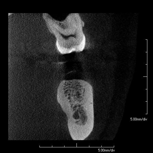
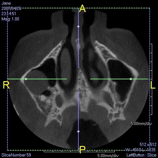
Sinus Lifts & Bone Grafts
- Highly accurate 3D images to determine if bone grafts or sinus lifts are needed
- Accurate information to diagnose maxillary sinus disorders
- The ability for a specialist to determine the 3D architecture of osseous defects
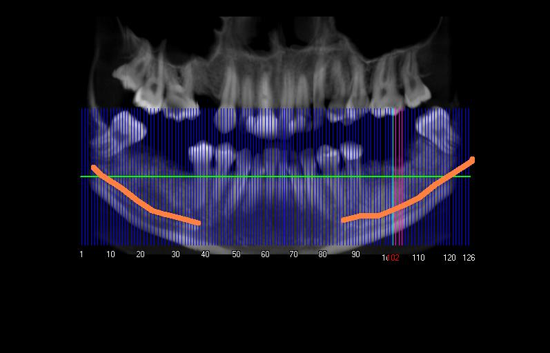
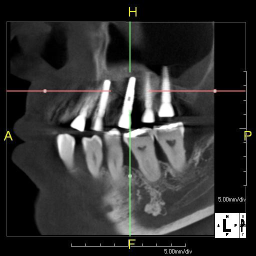
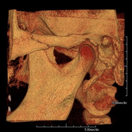
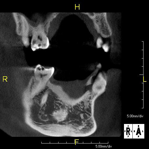
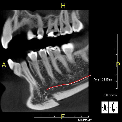
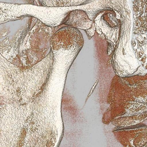
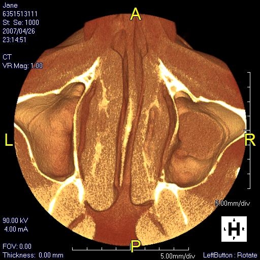
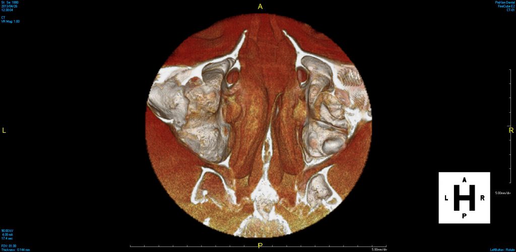
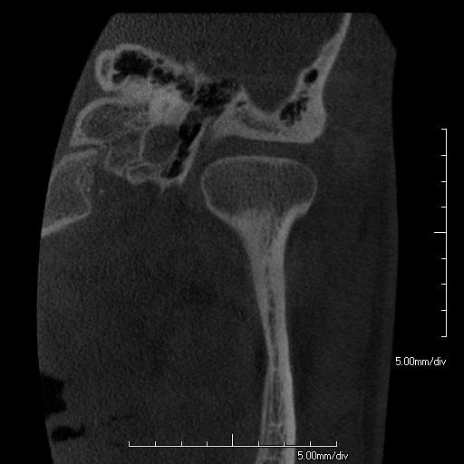
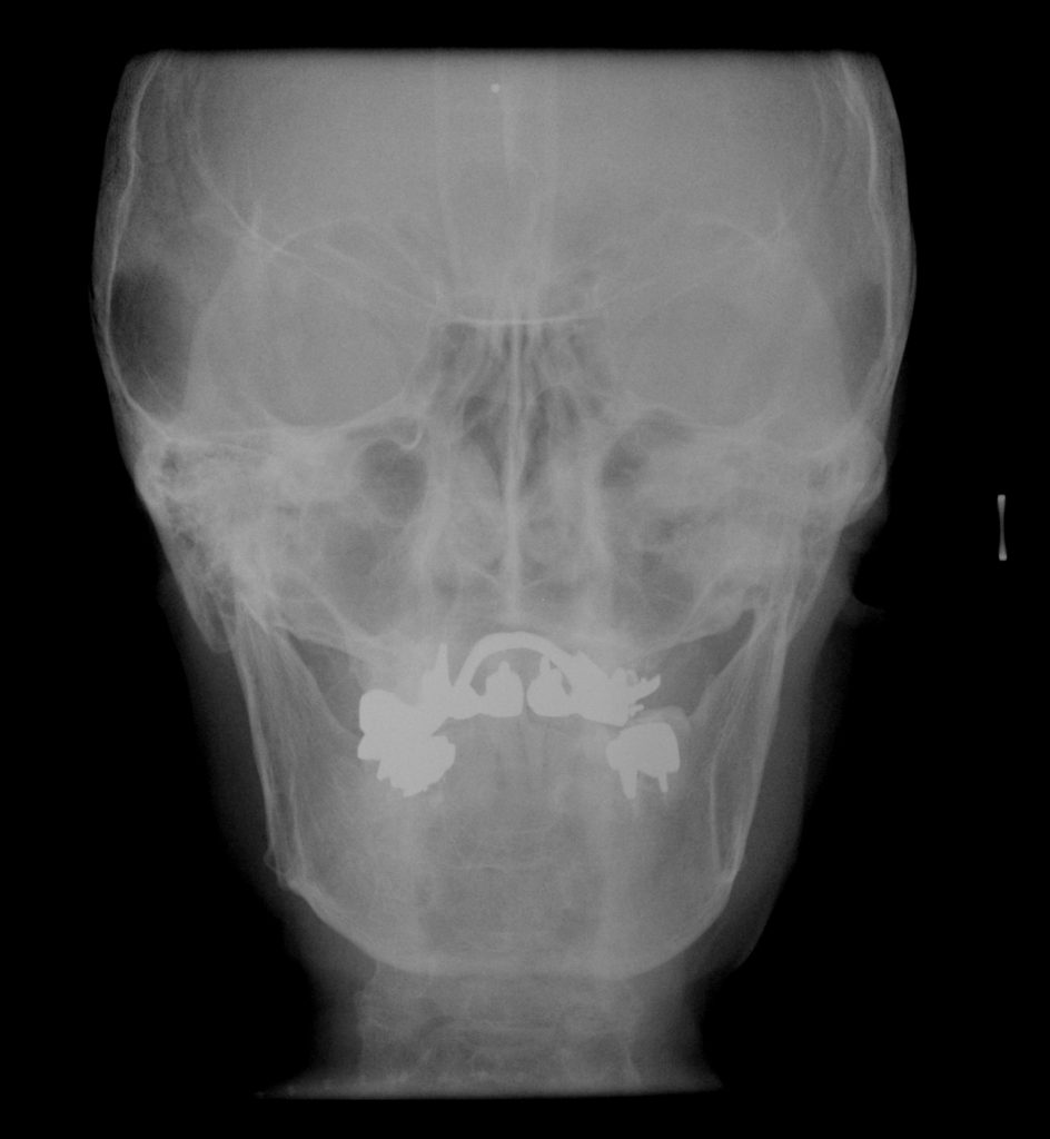
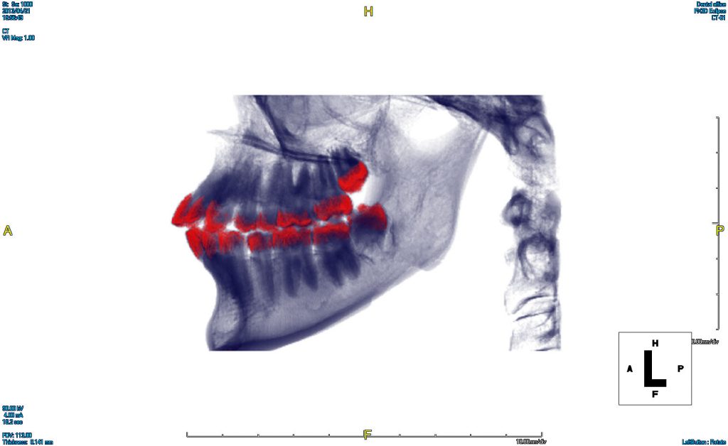
TMJ Studies
- Accurate assessment of the position, condition and contour of the Condyle, including the upper Ramus bone of the mandible
- Measurement tools that reveal exact distances between the Condyle and the bony Fossa, giving precise detail to aid the design and/or positioning for a corrective appliance
PreXion offers:
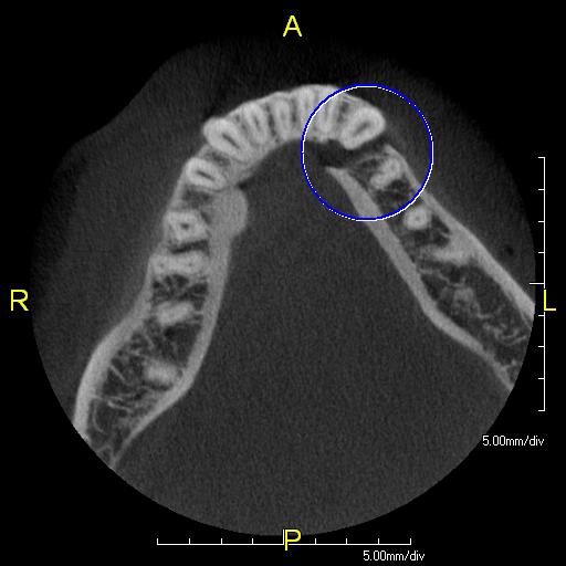
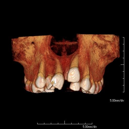
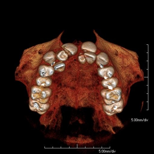
- Advantages of 1:1 geometry, including accurate measurements in three dimensions
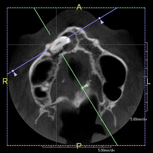
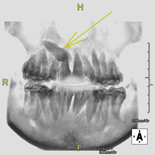
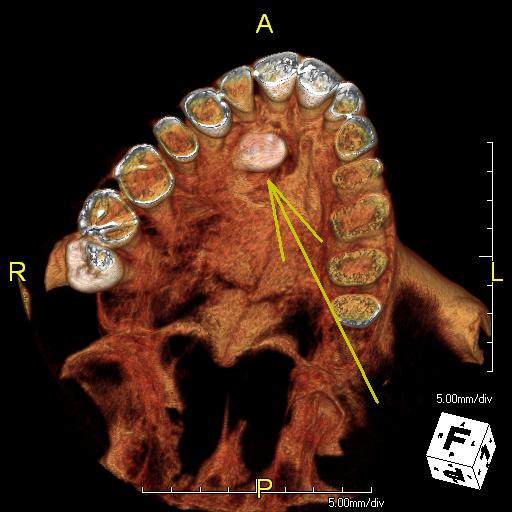
- Precise measurements of un-erupted teeth, bony dimensions
- Accurate localization of ectopic, impacted and supernumerary teeth
- Direct measurement of relative positions of teeth within skeletal components
- The ability to objectively assess asymmetries
- Three-dimensional views of airway and sinuses
- Confident pre-assessment of periodontal bone levels prior to orthodontic treatment without additional X-ray exposure
- Incidental endodontic findings that can alter final orthodontic planning
PreXion helps to:
- Easily export DICOM scans to third-party CAD/CAM software for merging data
- Integrate with all DICOM compliant third-party implant surgical planning software
- Plan implant cases more precisely with superior image clarity and resolution
- Streamline implant case workflow in your practice
- Increase case acceptance with patient education presentation tools
What Prosthodontists Are Saying…
“When we began looking at cone beam systems, medical CT scans were our gold standard. After several months of evaluating various systems, the PreXion 3D CBCT was the only one that provided us with the same high-quality images as a medical CT, with much less radiation. That truly was the deciding factor in our purchase decision.”
“Having the PreXion scanner in our practice has vastly improved our ability to treat patients efficiently and comprehensively. It allows the entire TEAM – specialists and general dentists – to have the most complete information for comprehensive treatment planning. PreXion is now an essential tool in our diagnostic tool kit.”
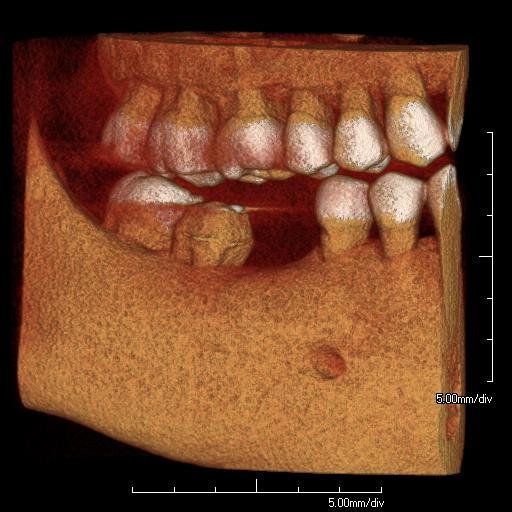
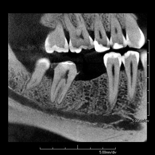
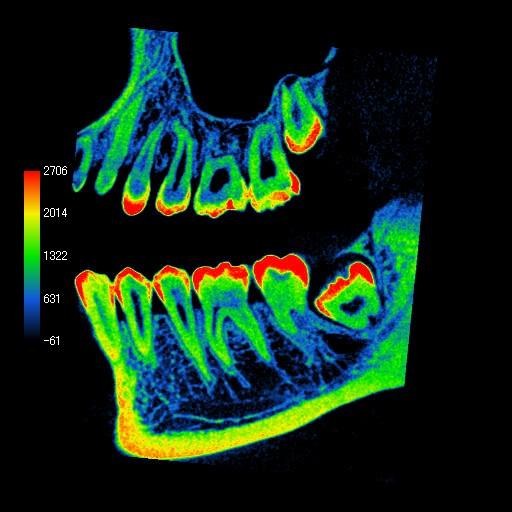
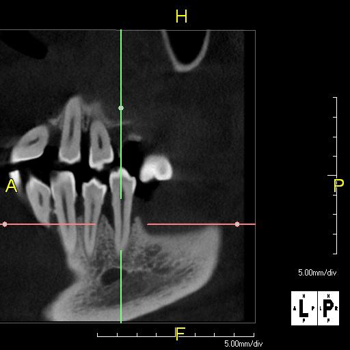
Periodontal defects can be visualized from any angle and the patient understanding of “pockets” is dramatically increased. The hygiene department will love the diagnostic value when treating deep defects.
PreXion helps to:
- Provide a sharper understanding of the bony defects relating to the surrounding anatomy
- Attain buccal and lingual measurement of osseous defects and periodontal lesions
- Assess PDL space
- See geometric relationships between the roots and furcation areas of mandibular first molars
- Determine bone level changes and periodontal defects
- Enable diagnosis of periapical pathosis as well as root resorption
- Measure the distance of the gingival margin to the facial bone crest
What Periodontists Are Saying…
“Without a question, this is the best tool I have in my arsenal to accurately diagnose and treat patients. The scanner’s operation is quick and simple, the software offers outstanding capability, and PreXion provides world-class, always-on customer support. The resolution of the PreXion 3D images and the ease of the software set the PreXion apart from any other scanner on the market. PreXion provides both the oral surgeon and me with a complete picture pre-surgery; I trust the data PreXion delivers each and every time.”
Joseph A. Sands, Jr., DDS
“I did quite a bit of research on CT systems before I made my decision to purchase the PreXion. I have been 100% satisfied with my decision. PreXion allows me to quickly and accurately diagnose and determine better treatment options for my patients. Prior to PreXion, I used a high quality 64 slice medical grade CT for my implant diagnosis and placement. I did not see any significant decrease in image quality when I changed to PreXion. Having the ability to illustrate and properly educate patients has genuinely helped us grow our practice. Likewise, PreXion’s sales and customer support is outstanding.”