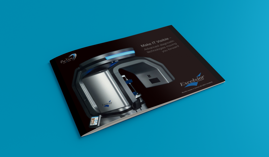
PreXion3D Excelsior CBCT Scanner
Bundled PreXion 3D Viewer Software
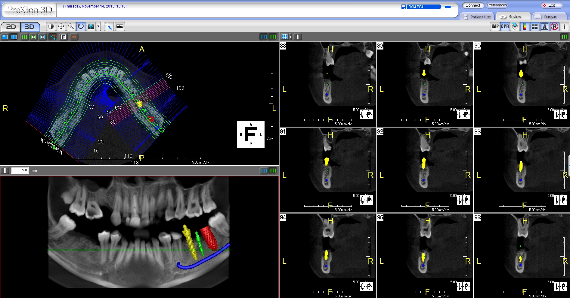
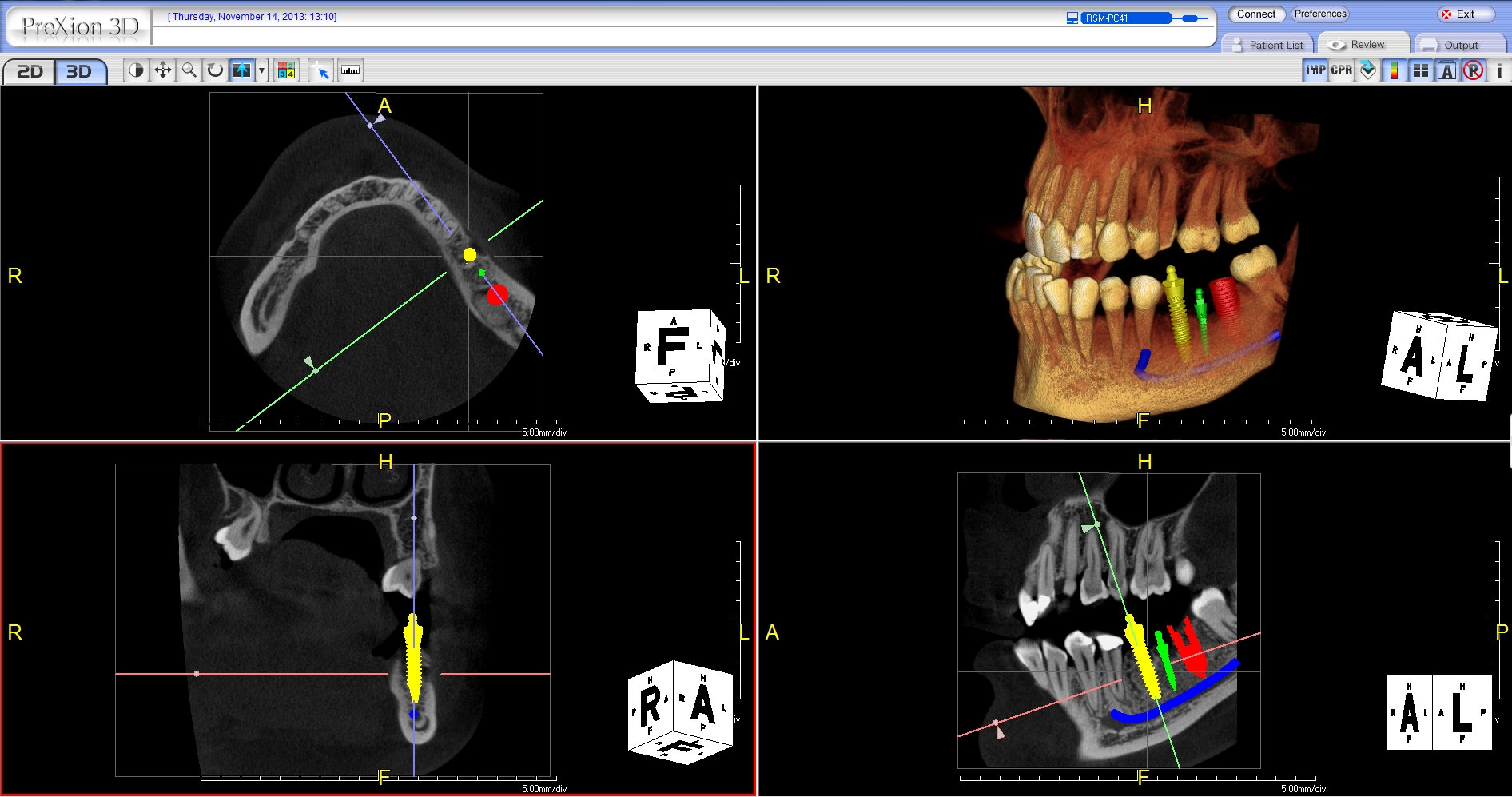
All PreXion3D CBCT scanners include The PreXion3D Viewer software at no additional charge. It contains easy modules for evaluating dedicated 2D Panoramic, 2D Cephalometric, and 3D images. PreXion provides the clinician with the most accurate assessment of the bone and surrounding anatomy while making 1:1 exact measurements. This assures optimal implant placement without the superimposition of tissue or projection distortion compared to conventional panoramic systems. PreXion3D reconstructs to DICOM 3.0 format and is compatible with major third-party surgical planning software systems.
• Better diagnose patients with more detail and clarity
• Present cases more confidently, increase acceptance
• Create the wow factor with patients
SOFTWARE FEATURES
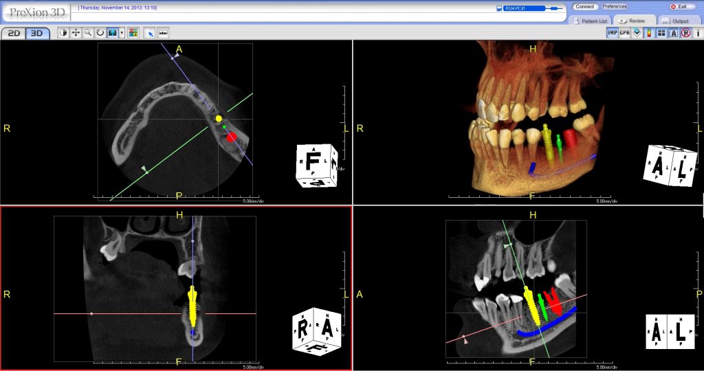
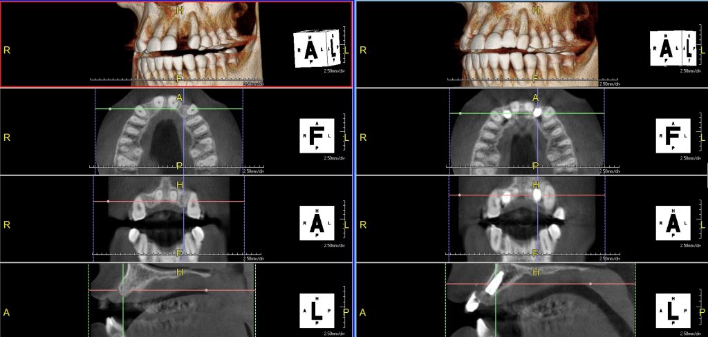
• Multi-data – Load multiple patient scans on a single screen. Synchronize pre- and post-operative scans and detect differences, slice-by-slice.
• Patient Education and Presentation – Quickly capture 3D animated video clips for patient education, case acceptance and lecture presentations. Increase case acceptance through better patient understanding.
• Collaborative Tools – Automatically save 3D image reports to MS Word template and attach to patient’s practice management record. Collaborate with referring dentists by burning a patient disc with sample viewer. Capture and email images quickly.
• Remote Access – Work on cases from home or a satellite office without long connectivity delays. Lead virtual online treatment planning meetings remotely with PreXion3D.
• Thin Client Server – PreXion3D CBCT scanners do not require computer hardware upgrades. It does not slow down network bandwidth like other CBCT scanners.
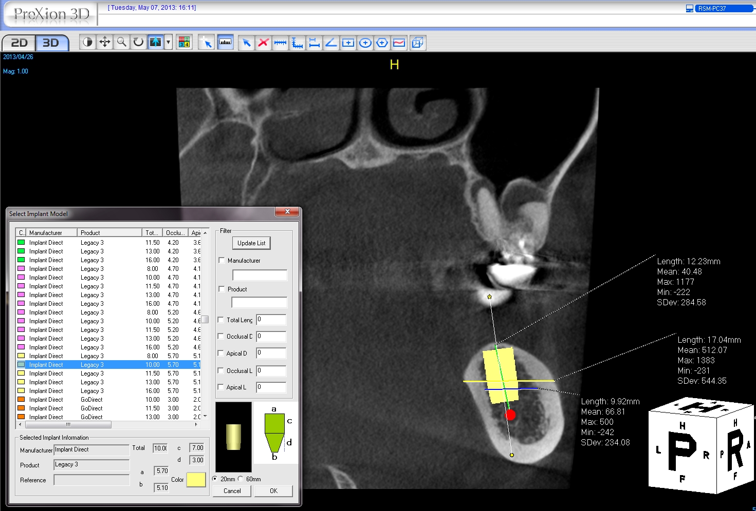
• Implant Library – Use our extensive library or customize your own library.
• Save Scenes – Save your case workup as a scene or create multiple saved scenes with a single scan.
• 3D Templates – Save time with over 20 pre-made 3D volume rendering templates or customize your own.
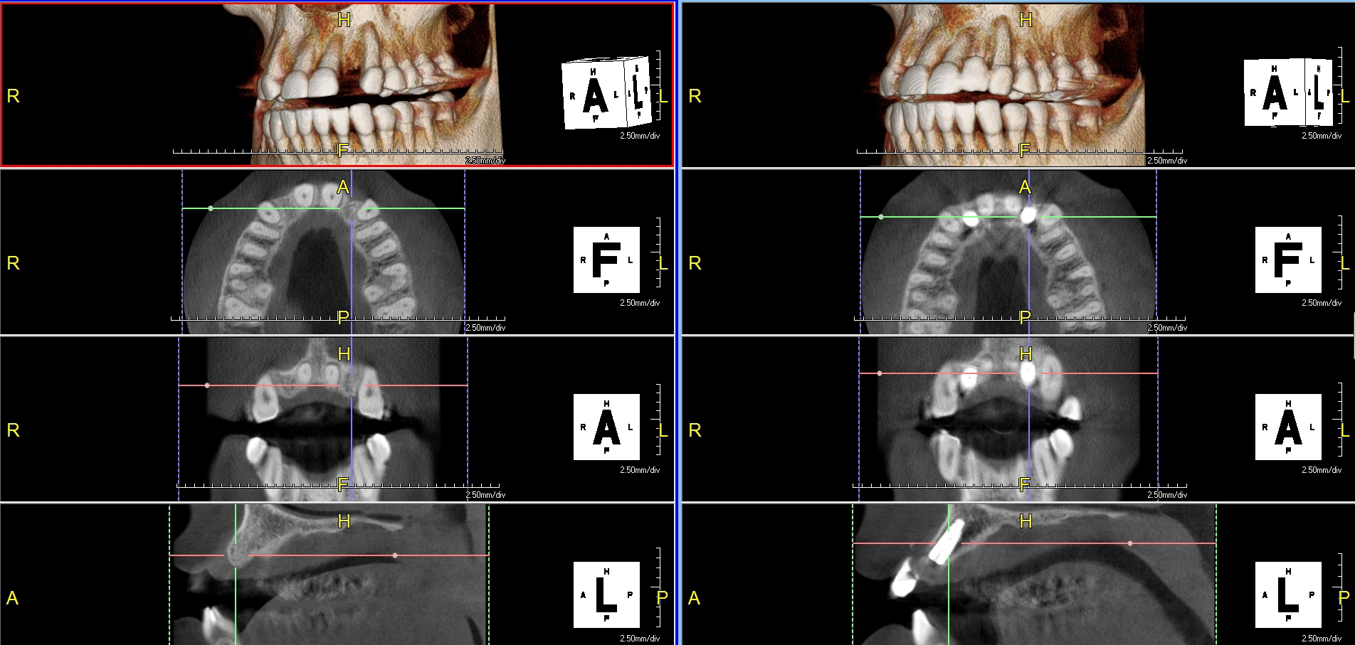
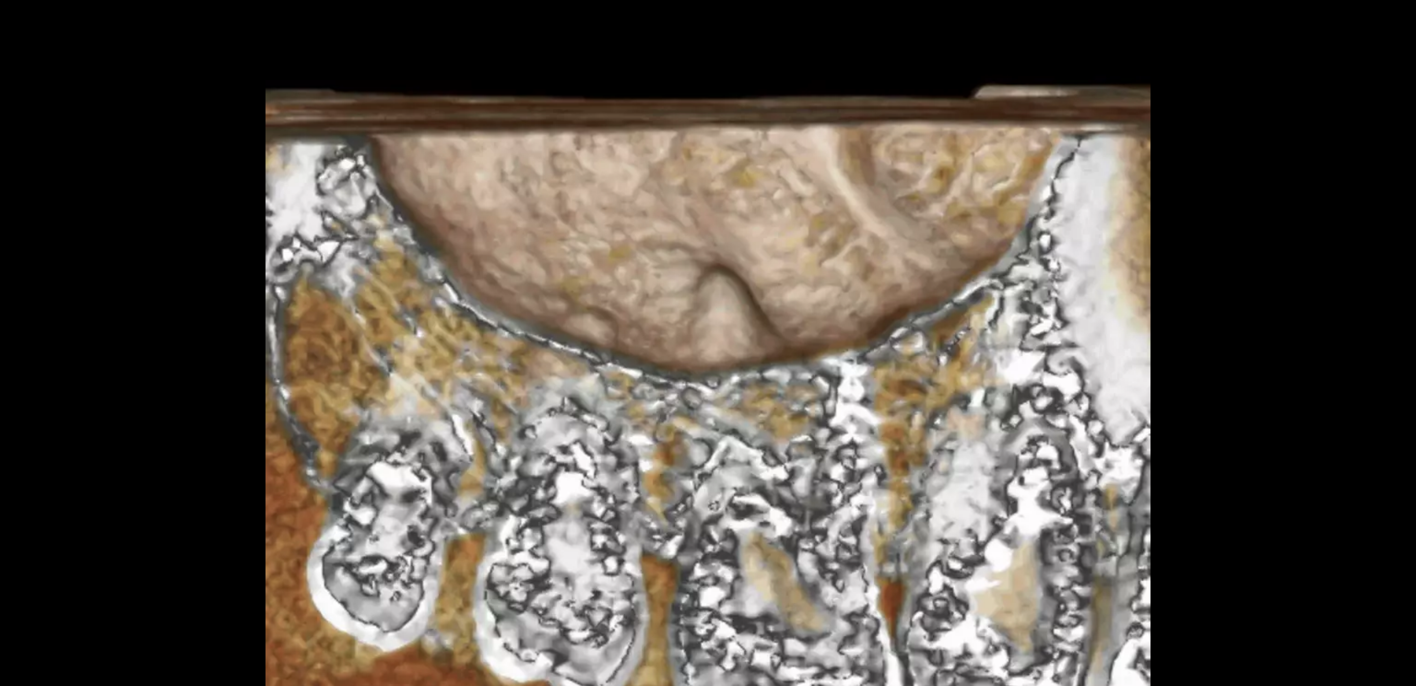
• Slab and Cutting – Slab Feature allows the clinician to see inside structures while rotating the 3D image. Cut away structures to see exactly what is pertinent to your study.
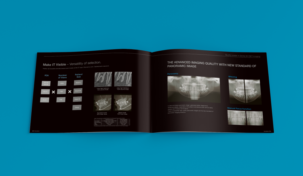
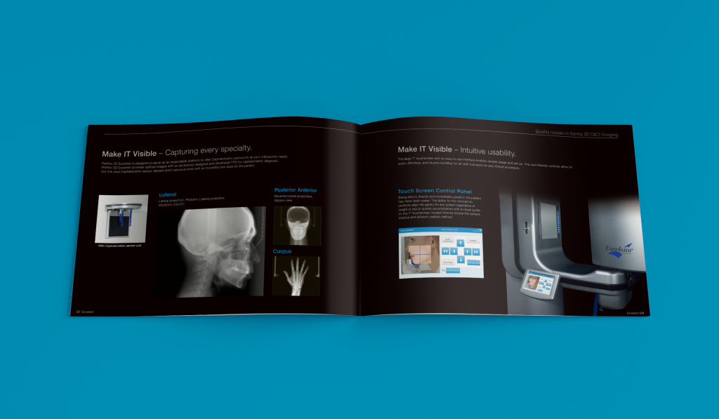
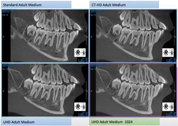
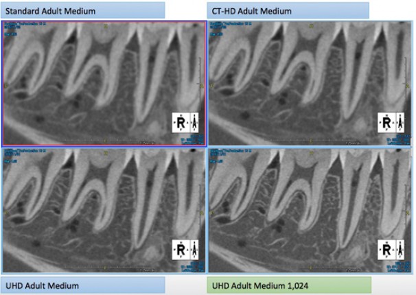
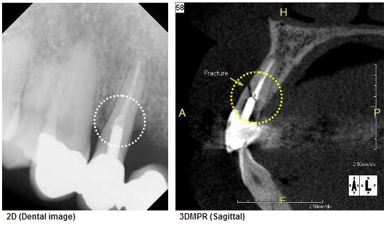
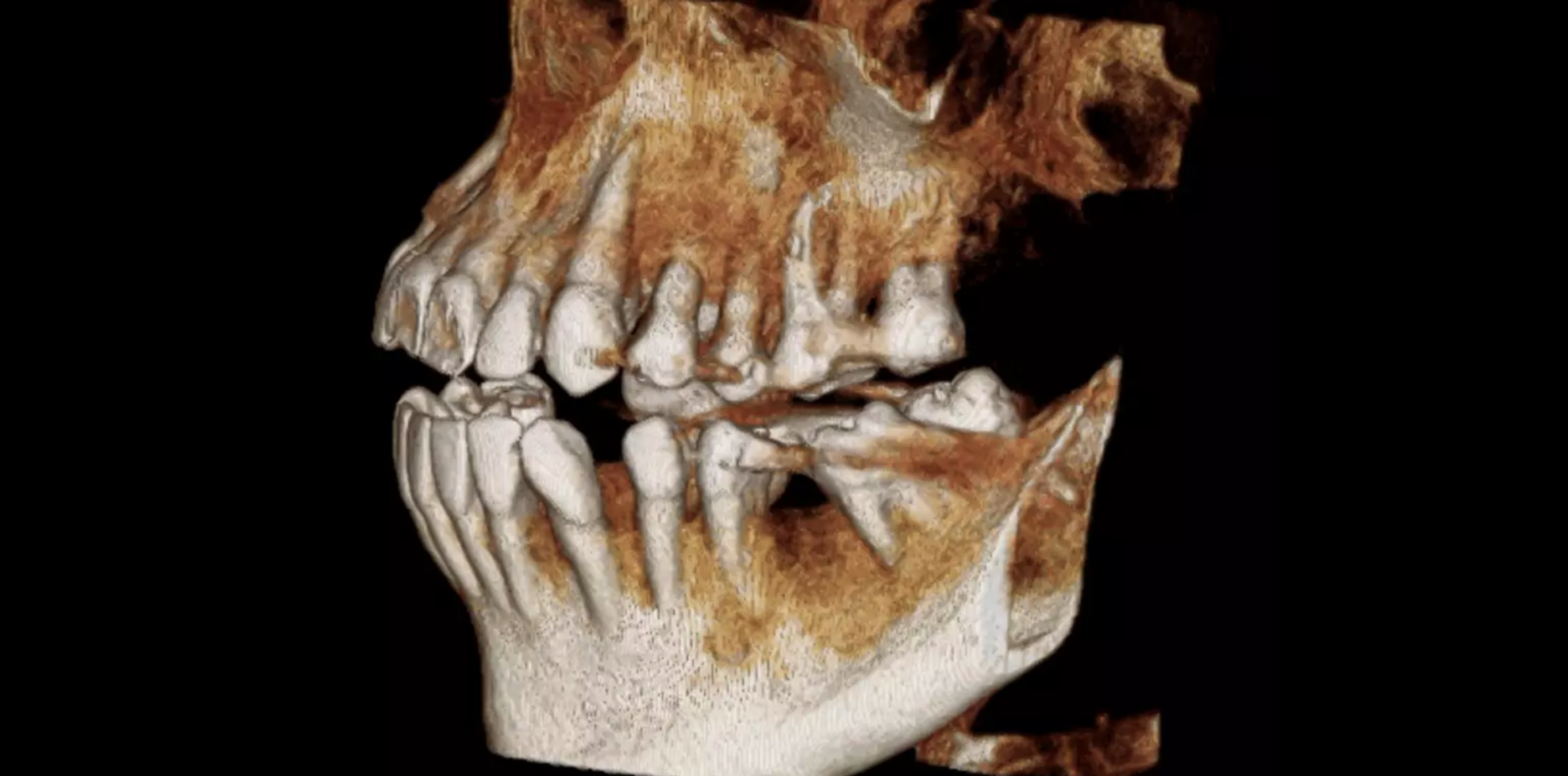
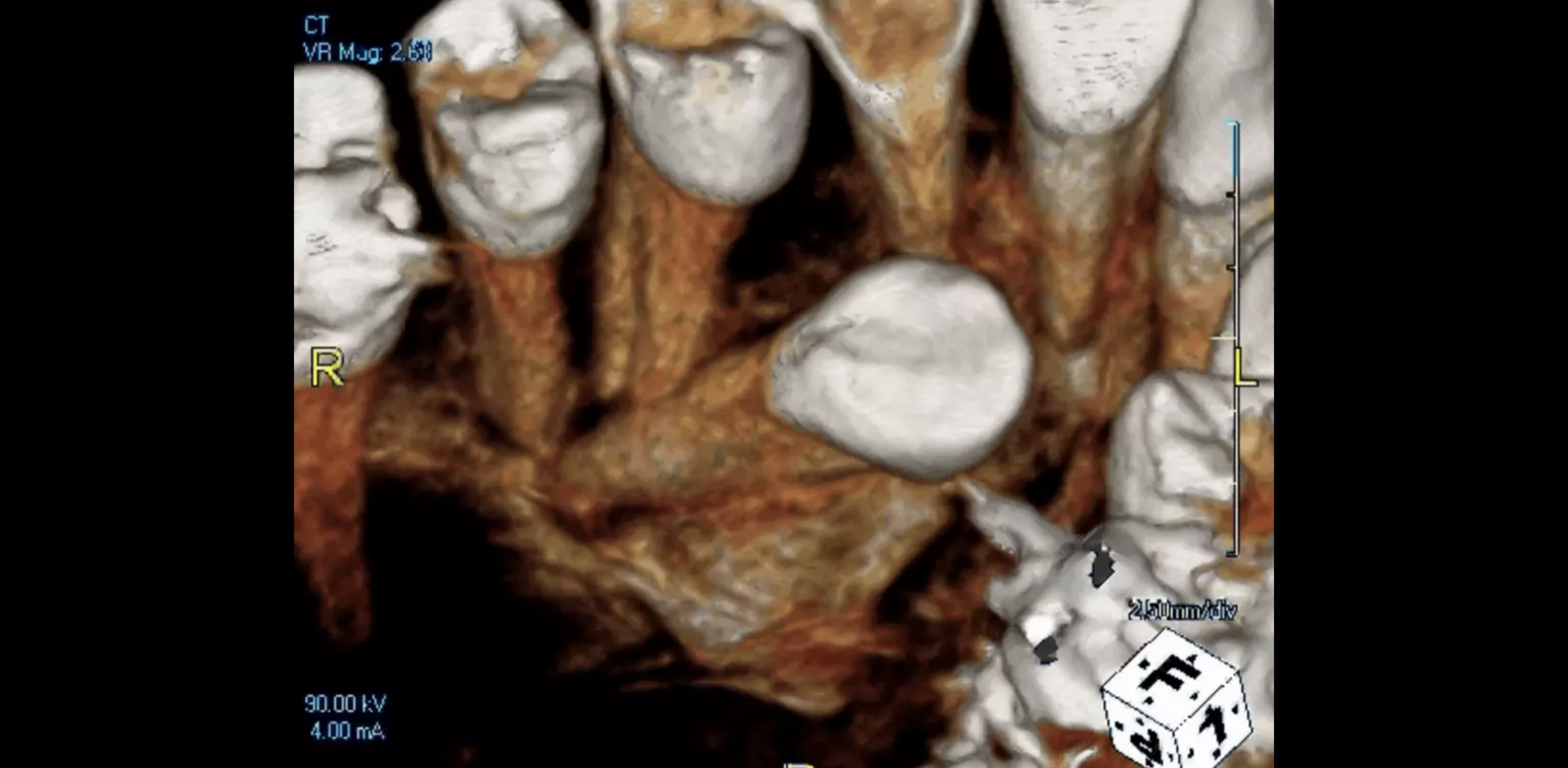
SOFTWARE IMAGES
CLINICAL APPLICATIONS
• Implant placement surgery
• Endodontics
• Periodontics
• Oral-maxillofacial surgery
• TMJ
• Pathology
• Impacted and supernumerary teeth
• DICOM export for implant surgical guides & CAD/CAM integration
• Airway analysis
HIGHLIGHTS
• Accurate 360-degree gantry rotation
• 260-1,024 projected views
• Dedicated 2D pan mode option
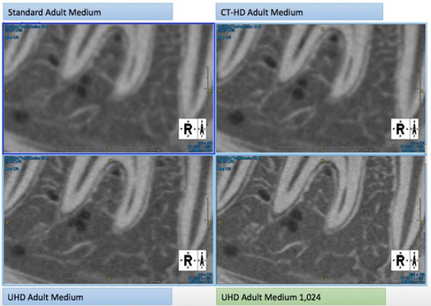
• Clearest detail with 0.3mm focal spot & 0.08-0.2mm voxel
SCAN MODE
| Standard Mode | 9.6 seconds |
| Rapid Mode (Lower Dose) | 5.2 seconds |
| High Definition Mode (HD) | 12.8 seconds |
| Ultra High Definition Mode (UHD) | 17.9 seconds, 23.6 seconds |
| Wide Mode | 9.6 seconds |
| CT-TMJ Mode | 5.2 seconds x 2 |
| CAD CAM Mode | 17.9 seconds |
| Panoramic Standard Mode | 10 seconds |
SPECIFICATIONS
| Device Type | CBCT+2D Panoramic + Cephalometric (Optional) |
| Field of View (DxH) | 150mm X 80mm; 100mm X 80mm; 100mm x 50mm; 50mm x 50mm; 150mm x 130mm (Optional) |
| CBCT Sensor | CsI FPD 16 bits, Cephalo FPD 14bits |
| X-ray Tube Voltage | 60–110kV |
| Focal Spot | 0.3mm x 0.3mm |
| Voxel Size | 0.08–0.2mm |
| Patient Position | Standing; wheel chair accessible |
| Included Computers | Single Console & Viewer Computer (Network Client is available) |
Product Videos
Testimonials
Testimonials
“When I was initially introduced to the PreXion CBCT technology, I was highly impressed with the image clarity. I was equally impressed with the support and training staff members that PreXion provided my practice. This made it easier for my staff to integrate this into my practice seamlessly.
Because the Midwest Implant Institute has taught post-doctoral cosmetic and dental implant procedures since 1986, we are very selective with whom we work with and the products and equipment we incorporate into our curriculum. Our attendees are amazed at the quality that brings PreXion 3-D imaging and case planning for restorative dentistry into their practices. The images make it easier to complete dental implant cases with precision, predictability and profitably.
PreXion’s CBCT system is not just for doctors who perform implant dentistry. PreXion technology for 3-D imaging provides a clinician the ability to diagnose endodontic and periodontal situations that can go undiagnosed with other traditional radiographic technology and techniques.”
